Technology
This doctor boasts a cutting-edge, fully-equipped digital in-house lab where every aspect of your dental care is seamlessly integrated under one roof. Experience the unparalleled advantage of minimized risk of cross-contamination, thanks to our state-of-the-art facilities. Say goodbye to discomfort with our digital workflow, eliminating the need for traditional impressions and the associated gag reflex. Witness the future of dentistry as you preview the final design before it’s even fabricated, allowing you to visualize your new smile with clarity and confidence. And best of all, enjoy the convenience of same-day dentistry, ensuring your dental needs are met efficiently and effectively. Welcome to a new era of dental care
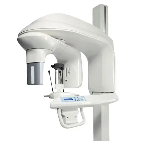
3D Cone Beam Imaging System
The current emerging standard of care in dentistry and dental implantology is the use of three dimensional x-ray studies. The 3-D images allow the doctor to collect the needed and highly valuable diagnostic information to best plan and deliver dental and surgical care.
Cone Beam imaging delivers quicker and easier image acquisition a typical scan takes only 20 seconds.
The patient benefits from less radiation as well as the comfortable, open environment. Aside from the physical comfort of this system, the doctor can share a visual diagnosis with patients, making them more comfortable with their treatment plan and actively participate in their care. The speed of the scan and the immediate results allows the doctor and patient to better communicate the aspects of a case.
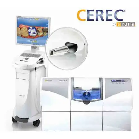
CEREC 1 Visit Tooth Restoration
CEREC Technology allows us to provide our patients with Same Day Restorations.
No more messy impression materials or hassle with annoying temporaries! CAD/CAM Technology offers our patients the opportunity to have a beautiful restoration designed, fabricated and delivered in a single appointment! Here’s how:
Using, CEREC Technology, we are able to take a photo scan of your tooth with a special laser camera that safely captures the anatomy of the prepared tooth and renders it as a 3-dimensional image.
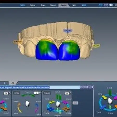
We then customizes your crown, inlay or onlay using your digital “impression” and 3D imaging software while you relax with a magazine or music headphones.
After the doctor approves the final design, the CEREC milling system employs CAD/CAM technology to shape your restoration from a high-strength, metal-free ceramic or composite block in a shade to match your smile. This eliminates the repeat visit after sending the impression off to a lab, as this is now able to be done right in the office while you are in the chair!
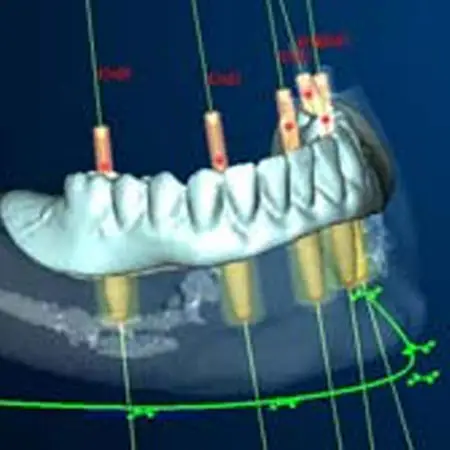
Computer Guided Implant Surgery
We use state-of-the-art technology including Computer Guided Implant Surgery to ensure the optimum results in patient care.
In the past radiographic images used to place implants were two dimensional providing a reasonable estimate of bone height. Often bone width was not determined until the bone was exposed during surgery sometimes resulting in compromised placement of the implant.
Computer Guided Implant Surgery gives very detailed 3-D visualizations and coupled with the appropriate software allows us to place implants with a level of precision that was unattainable several years ago. In addition, the surgical procedures can often be accomplished in a much more conservative procedure which involves greatly reduced discomfort, less treatment time and a more accurate final outcome.
Computer Guided Implant placement is the safest and most precise way to place dental implants.
DIAGNOdent Laser Cavity Detection
DIAGNOdent is an advanced laser technology that helps us find tooth decay more accurately and reliably in its earliest stages, before it causes more widespread damage. We use it along with visual examination, dental explorers, and x-rays. DIAGNOdent helps us locate decay that’s hidden inside the tooth where visual examination, explorers, and x-rays can’t find it. The DIAGNOdent hand piece scans your teeth with harmless pulses of laser light. When the laser reaches decay under the surface of the tooth, the decay emits a fluorescent light. This fluorescent light bounces back to the sensor, and is translated into an audible signal and a digital readout. In general, the higher the number, the greater the amount of decay in the tooth.
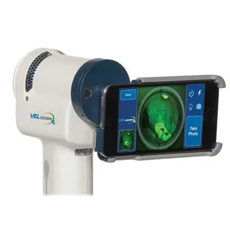
Early Detection for Oral Cancer
It is a shocking statistic that one American dies each hour because of oral cancer. This mortality rate has not changed in 40 years! But the upside is that when oral cancer is detected in its early stages, 90% of these patients can be cured.
We have the VELscope™ available for our patients for screening and early detection of this life-threatening problem. It is an FDA-approved specially designed light. Immediately, any unusual tissue can be identified. This is a quick, easy, and painless exam that could save your life!
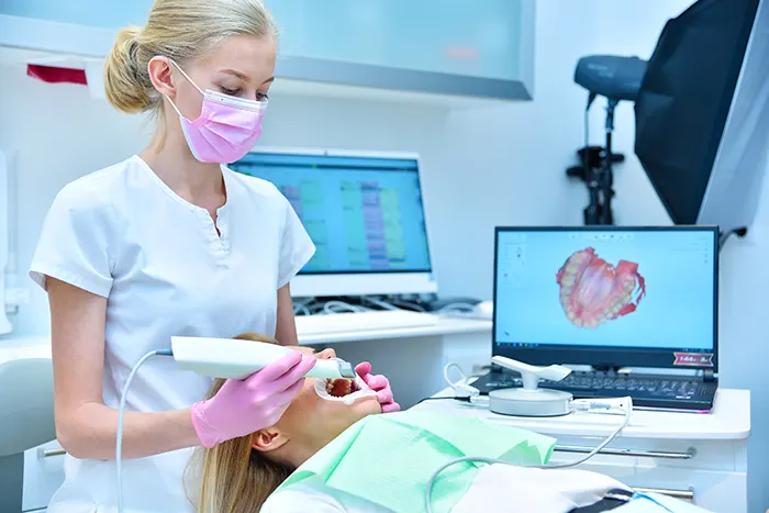
Intra-Oral Camera Lets You See for Yourself
Through technology, our patients can work together with our dentists to have healthy gums and beautiful teeth. With the intra-oral camera, you can see for yourself what work needs to be done on what teeth. This amazing miniature video camera produces clear and accurate pictures of all your teeth. You will enjoy making informed decisions together with our doctors, creating a bond of teamwork.
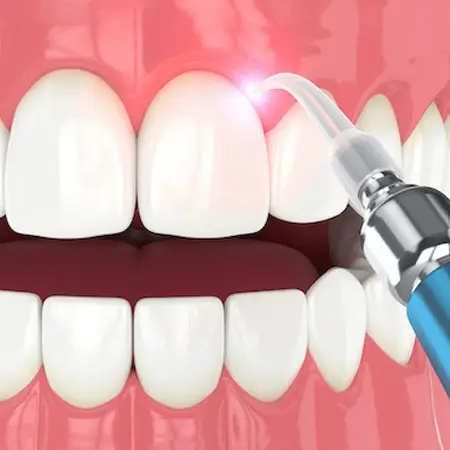
Laser Dentistry
Laser Dentistry offers you the opportunity to have a safe, anxiety free dental experience, without drills and scalpels, and in most cases, without anesthesia. The laser acts as a cutting instrument or a vaporizer of tissue that it comes in contact with in surgical and dental procedures.
Laser Dentistry uses laser energy and a gentle spray of water to perform a wide range of dental procedures – without the heat, vibration and pressure associated with the dental drill. The laser beam sterilizes the affected area and seals off blood vessels, which minimizes the chance of infection or bleeding. Patients are also much more comfortable during and after treatment with laser therapy
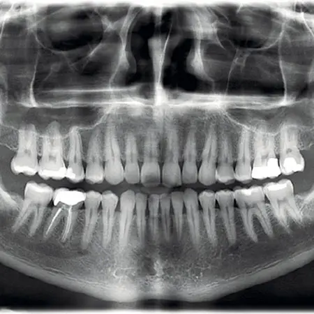
Low Dose Digital X-Rays
Dental x-rays (Radiographs) are essential, preventative, diagnostic tools that provide valuable information not visible during a regular dental exam. Digital Radiography is a type of X-ray imaging that uses digital X-ray sensors to replace traditional photographic X-ray film, producing enhanced computer images of teeth, gums, and other oral structures and conditions. This allows the doctors to get a much better view of your teeth and any potential dental conditions. The digital imaging software obtains a number of different views of your teeth to gain the best understanding of your situation and develop the most appropriate course of management.
Digital sensors are more responsive than film so that much less radiation is required to produce a digital image.
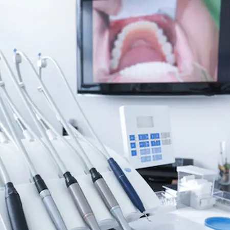
Rotary Endodontics
A root canal is a treatment used to repair and save a tooth that is badly decayed or becomes infected. During a root canal procedure, the nerve and pulp are removed and the inside of the tooth is cleaned and sealed. Without treatment, the tissue surrounding the tooth will become infected and abscesses may form.
Rotary Endodontics is the procedure in which a power driven instrument is used for root canal treatments. The use of power driven instruments allows for more accurate cleaning and shaping of your root canal, thus allowing it to be filled easier and with more predictable results. This technology also dramatically reduces post-operative sensitivity, time spent in the dental chair and discomfort compared to conventional endodontic treatment.
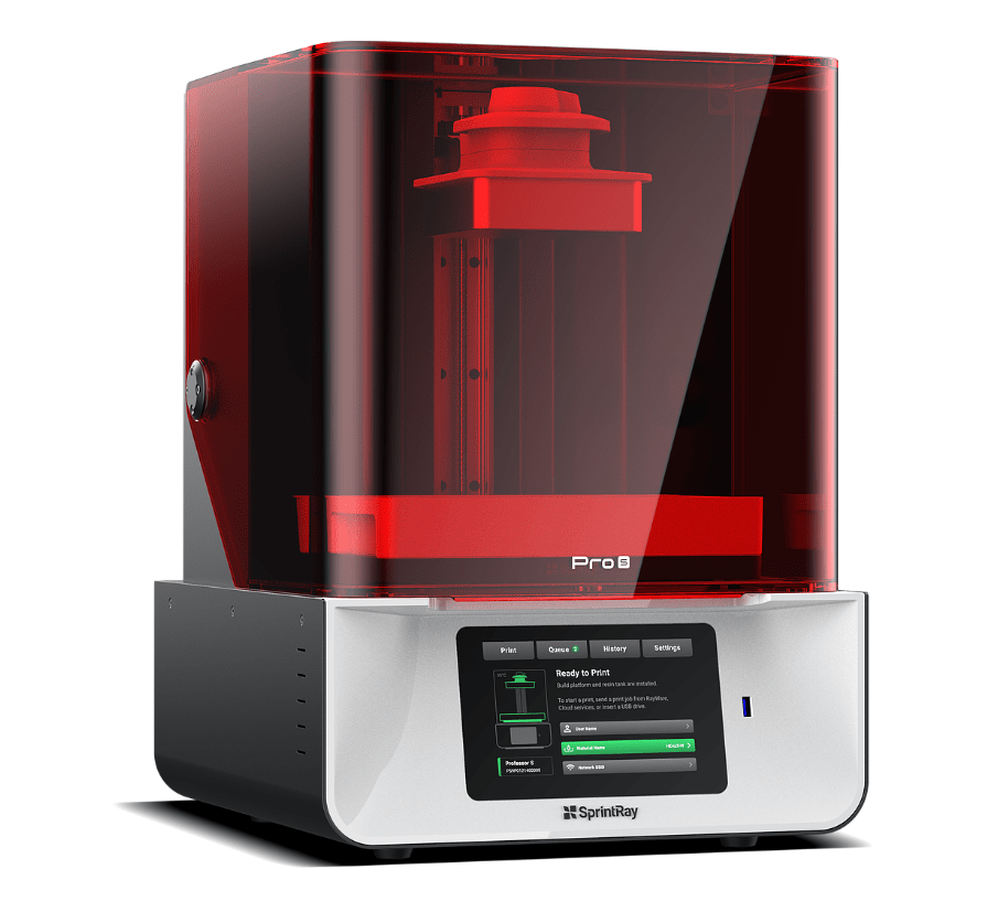
Sprintray Digital Printer
With our Sprintray Digital 3D Printer, we have the ability to print models, surgical guides, and other useful tools in our office. This precise, flexible device can improve the quality of dentistry we can offer. For example, we can take digital scans of your smile and print a surgical guide, making procedures like dental implant surgery safer and more accurate.

